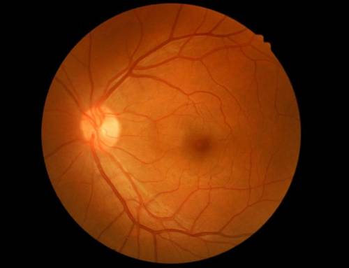
Ocular hypertension means that the internal pressure of the eye is higher than normal. Ocular hypertension is not a disease, but it can lead to serious illnesses such as glaucoma, the “thief of sight”.
Intraocular pressure is the result of the pressure exerted by the liquids inside the eye against the walls of the eyeball. When there is no balance between the production and absorption of these liquids, an increase in pressure can occur, in other words ocular hypertension.
For this reason it is important that if we have a family history and if the ophthalmologist detects it in a routine examination, that we follow his advice and comply with the treatment for ocular hypertension.
How to measure eye pressure
The main drawback when measuring eye pressure is that we do not have access to the inside of the eyeball. There is no valve or outlet where we can “connect” a pressure gauge, so other methods of measurement are used.
The eye pressure is taken on the walls of the eyeball, usually on the cornea which is the most accessible. A tonometer is used for this purpose, and as a result we must have a 16 mmHg (millimetre of mercury).
The fact is that we do not all have the same corneal thickness, so the calculation is not so simple and requires a corneal pachymetry. The pachymetry allows us to know the thickness of the corneal wall. This data is important because when a cornea is thick we can obtain as a result a high false pressure and vice versa, when it is thin we can obtain a low false pressure.
We give you a very clear example so that you can see the importance of carrying out a pachymetry test. If 16 mmHg is the accepted mean value, it turns out that the corneal thickness can cause a deviation in the calculation of up to 10 mmHg.
The balance between production and reabsorption of the aqueous humour is the main factor that determines the level of intraocular pressure.

Leave A Comment